Anatomy Muscles Pelvis - Your Pelvic Floor Muscles Why You Should Care Our Fit Family Life. It is composed of three separate paired muscles; The structure of the pelvis supports the contents of the abdomen while also helping to transfer the weight from the spine to the lower limbs. The pelvic floor muscles include; A proper kegel exercise means a full contraction and relaxation of the pc muscle. The pelvic brim involves the first sacral segment, the iliac and pubis portion, but not the ischium.
▪these four bones are connected by four joints and lined by four muscles. Time to solidify your knowledge on the anatomy of the. The pubococcygeus (pc) muscle is the muscle that runs the show in pelvic floor health. Small and deep muscles which mainly externally rotate the thigh at the hip joint and stabilize the pelvis. A proper kegel exercise means a full contraction and relaxation of the pc muscle.
Muscles Of The Pelvic Floor Anatomy And Function Kenhub from thumbor.kenhub.com They are responsible for bending and adducting the thigh. The function of the pelvic floor is to help assist with child birth, prevent incontinence and support organs within the pelvis. The floor of the pelvis is formed by the two muscles named levator ani and coccygeus. These muscles have attachments to the pelvis as follows: The levator ani muscles consist of three. The hip joint is one of the most flexible joints in the entire human body. Ligaments, tendons, and muscles play an important role in the function of the hip. Muscles that attach from the pelvis to the trunk and cross the lumbosacral joint muscles that attach from the pelvis to the thigh/leg and cross the hip joint pelvic floor muscles that are located wholly within the pelvis
These are the piriformis, obturator internus, obturator externus, gemellus superior, gemellus inferior, and quadratus femoris.
Rectus femoris muscle, one of the quadriceps muscles on the front of your thigh. Then we'll look at the complex sheet of muscles, collectively called the pelvic diaphragm, which form the floor of the pelvic cavity. •sacrumandcoccyx, which form the posterior wall. The function of the pelvic floor is to help assist with child birth, prevent incontinence and support organs within the pelvis. Time to solidify your knowledge on the anatomy of the. These two muscles join each other and then attach to the lesser trochanter. The four groups are the anterior group, the posterior group, adductor group. This mri male pelvis axial cross sectional anatomy tool is absolutely free to use. The iliopsoas muscle consists of the iliac muscle, which comes from the inner surface of the ilium in the pelvis, and the psoas muscle, which originates from the vertebral column. They are responsible for bending and adducting the thigh. The pelvis's frame is made up of the bones of the pelvis, which connect the axial skeleton to the femurs, and therefore acts in weight bearing of the upper body. These are the piriformis, obturator internus, obturator externus, gemellus superior, gemellus inferior, and quadratus femoris. The muscles within the pelvis may be divided into two groups:
Piriformis and obturator internus are both hip rotator muscles, which arise within the pelvis, and pass outward through the sciatic foramina. •two hip bones, which form the anterior and lateral walls. We'll look at these structures first in a male specimen. They form a large sheet of skeletal muscle that is thicker in some areas than in others. The pelvic brim involves the first sacral segment, the iliac and pubis portion, but not the ischium.
Pelvic Muscle Anatomy Anatomy Drawing Diagram from i0.wp.com The pelvic floor muscles provide foundational support for the intestines and bladder. ▪the bony pelvis is composed of four bones: Arcus tendineus levator ani and the ischial spine A proper kegel exercise means a full contraction and relaxation of the pc muscle. The muscles of the pelvis and hip control the vast range of movement of the legs and torso. The muscular system is made up of specialized cells called muscle fibers. •sacrumandcoccyx, which form the posterior wall. Piriformis and obturator internus are both hip rotator muscles, which arise within the pelvis, and pass outward through the sciatic foramina.
The muscles of the pelvic floor are collectively referred to as the levator ani and coccygeus muscles.
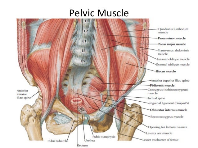
▪these four bones are connected by four joints and lined by four muscles. Rectus femoris muscle, one of the quadriceps muscles on the front of your thigh. This mri male pelvis axial cross sectional anatomy tool is absolutely free to use. These muscles, including the gluteus maximus and the hamstrings, extend the thigh at the hip in support of the body's weight and propulsion. Arcus tendineus levator ani and the ischial spine ▪the bony pelvis is composed of four bones: The pelvic inlet is delineated by a bone crest that defines its limit (the pelvic brim), which later refers to the promontory of the sacrum. The muscles of the pelvic floor are collectively referred to as the levator ani and coccygeus muscles. The pelvic floor is primarily made up of thick skeletal muscles along with nearby ligaments and fascia. The levator ani muscles are the largest group of muscles in the pelvis. Psoas consists of a pair of deep muscles (psoas major and iliacus) located on each side of the pelvis in the abdomen. These are the piriformis, obturator internus, obturator externus, gemellus superior, gemellus inferior, and quadratus femoris. The function of the pelvic floor is to help assist with child birth, prevent incontinence and support organs within the pelvis.
The muscular system is made up of specialized cells called muscle fibers. They are responsible for bending and adducting the thigh. These two muscles join each other and then attach to the lesser trochanter. These muscles have attachments to the pelvis as follows: However, often they will do more than one movement, assisting another muscle.
Pelvic Muscles Anatomy Anatomy Drawing Diagram from image.slidesharecdn.com The floor of the pelvis is formed by the two muscles named levator ani and coccygeus. Rectus femoris muscle, one of the quadriceps muscles on the front of your thigh. The hip joint is one of the most flexible joints in the entire human body. The pelvis consists of the sacrum, the coccyx, the ischium, the ilium, and the pubis. These muscles can be grouped based upon their location and function. These muscles origin in continuity from the body of the pubis, along a tendinous arch over the obturator internus fascia, and the ischial spine. Attached to the pelvis are muscles of the buttocks, the lower back, and the thighs. (1) the obturator internus and the piriformis, which are muscles of the lower extremity, and will be described with these (pages 476 and 477);
These muscles all serve as adductors of the thigh, but also serve as important stabilizers of the pelvis and work to maintain balance of the pelvis on the lower limb during gait.
They are also known as the inner hip muscles and deep external rotators. The muscles within the pelvis may be divided into two groups: They form a large sheet of skeletal muscle that is thicker in some areas than in others. They are responsible for bending and adducting the thigh. The muscles of the pelvic floor are collectively referred to as the levator ani and coccygeus muscles. However, often they will do more than one movement, assisting another muscle. The levator ani is a broad sheet of muscle. The many muscles of the hip provide movement, strength, and stability to the hip joint and the bones of the hip and thigh. It is composed of three separate paired muscles; To support the abdominal and pelvic viscera They have several functions, including helping to support the pelvic organs. Each of the hip muscles will have a main function, to produce a specific movement. Large ligaments, tendons, and muscles around the hip joint hold the bones (ball and socket) in place and keep it from dislocating.
Share :
Post a Comment
for "Anatomy Muscles Pelvis - Your Pelvic Floor Muscles Why You Should Care Our Fit Family Life"


:background_color(FFFFFF):format(jpeg)/images/library/12123/841_842_843_m_transversus_perinei_m_bulbospongiosus_m_ischiocavernosus_m_sphincter_ani_m_sphincter_urethrae.png&description=Anatomy Muscles Pelvis - Your Pelvic Floor Muscles Why You Should Care Our Fit Family Life)
Post a Comment for "Anatomy Muscles Pelvis - Your Pelvic Floor Muscles Why You Should Care Our Fit Family Life"